
Layer Visualisation in Recycled PET Sheets
The incorporation of recycled plastic material for packaging is now an established reality in the packaging sector. Legislation mandates this, and in Spain, starting in 2025, PET packaging must contain at least 25% recycled material, according to Royal Decree 1055/2022. However, despite the widespread use of recycled material, some applications that require monitoring and quantifying its presence present difficulties.
This is the case with three-layer coextruded PET sheets with an A/B/A structure for tray thermoforming, where A is virgin PET and the intermediate layer B is recycled PET. These sheets present a problem when attempting to verify the presence of this structure and even the thickness ratio. The transparency of the materials used and the coextrusion process itself make it impossible to verify the multilayer structure using conventional methods. A cross-section of the film taken with a microtome and viewed under a light microscope shows that there is no distinction between the three layers in the image obtained (see photo 1).
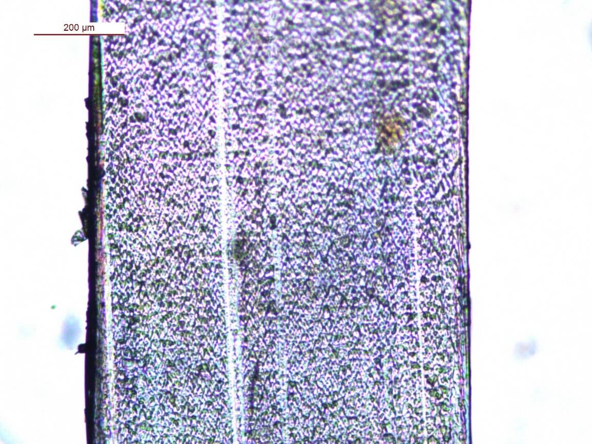
Figure 1. Transmission micrograph at room temperature, 5x objective
Since sample preparation and viewing using light microscopy are a quick way to observe the layer distribution in a multilayer film, it is frustrating not to be able to use this technique in these types of samples.
To address this issue, several tests have been conducted in the AIMPLAS Characterization laboratories using different samples of this type of film, with cross-sectional views taken at various temperatures. Using a special accessory for the light microscope, a heating plate, a controlled temperature scan can be applied from ambient to higher temperatures (no more than 180°C) over the film cross-section, which can be observed while the scan is in progress. The cut PET/RPET/PET sheet will be heated in a controlled manner and will undergo the typical transitions of this material.
The purpose of observing the heating of the sample is to determine whether the two types of PET exhibit different behavior, revealing any optical changes that could be used to differentiate them. Viewing the temperature sweep provides images of the different stages of PET heating, and it is when cold crystallization occurs that the trilayer structure is revealed due to the different nature of the two types of PET used to manufacture these sheets (see photo 2).
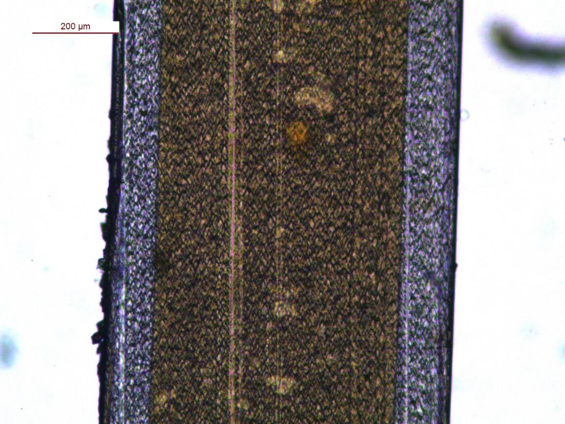
Figure 2. Transmission micrograph at 140°C, 5x objective
As can be seen, the trilayer structure is clearly distinguishable at 140°C, making it possible to verify the presence of two different materials and even to check the thickness ratio.
In this way, AIMPLAS provides a solution to the monitoring problem presented by this type of film. This method can be useful both for production control and for evaluating the quality of manufactured films.